The application value of three-dimensional ultrasonic rotary cutting in the diagnosis of liver tumor
The disease classification is shown in Table 1. 17 cases of metastatic liver cancer were diagnosed according to the history of primary tumor and multiple intrahepatic tumors. 28 cases of hepatic hemangioma were diagnosed according to years of follow-up and typical ultrasound signs, and the other cases were confirmed according to surgical pathology or biopsy.
Table 1 85 cases of liver tumors, two-dimensional image echo, statistics, disease cases, echoes, high echo, low echo, mixed echo, echo, hepatic hemangioma, primary liver cancer, metastatic liver cancer, hilar cholangiocarcinoma, hepatic leiomyoma, total method, 1.2 method, three-dimensional ultrasound The workstation is developed by Beijing Zhongke Hengye Technology Co., Ltd., and connected with Sequia512 ultrasonic machine. The frequency of the convex array probe is 3.0~5.0MHz. Image acquisition method: Firstly, the conventional two-dimensional scan of the tumor to be examined is performed (Table 1), and the best selection is made. The two-dimensional image is stored in a photograph, and then the image is uniformly collected by a fan-shaped or parallel free scanning method, and the image is transmitted to the three-dimensional ultrasonic workstation. After the image is collected, the tumor is centered on the dynamic rotary cutting, and the internal structure of the tumor of different cut surfaces is observed. The relationship between the surrounding tissues, selecting a cut surface similar to a two-dimensional image to store photographs, facilitates comparison of two-dimensional and three-dimensional images.
A. Two-dimensional ultrasound image showing that the tumor is close to the echo of the same echo 2 results. Eighty-five cases of liver tumors were successfully performed by three-dimensional ultrasound dynamic rotation cutting method, which clearly showed the shape, structure and echo of different sections in the tumor and the tumor edge, and simultaneously showed The relationship between the tumor and the surrounding tissue and adjacent organs provides more and more diagnostic information than the two-dimensional ultrasound, such as the hypoechoic halo at the edge of the liver cancer, partly composed of edema, and partly composed of blood vessels. Through dynamic rotation, the display of liver tumors is more realistic, the stereoscopic effect is strong, and the spatial position is clear, just like seeing the tumor entity specimens. () The echoes of the two-dimensional images of liver tumors are shown in Table 1, 31 strong echogenic lesions and 21 mixed echoes. The three-dimensional image of the lesion is significantly better than the two-dimensional image, which clearly shows the boundary of the tumor and the characteristics of the internal echo (see the third). The three-dimensional image of the 23 hypoechoic lesions is also superior to the two-dimensional image, which clearly shows the boundary of the tumor. It also clearly shows the characteristics of internal hypoechoic, but if the echo is too low, it gives a feeling of tissue defect or void (see 3); 10 cases of echoes or echoes close to the echo are very blurred. However, three-dimensional ultrasound can well show the boundary of the tumor and the characteristics of the internal echo, which is obviously superior to the two-dimensional ultrasound image (see the third) B-dimensional ultrasound image. The image shows the surface shape and internal structure of the tumor more clearly. Specimens like liver cancer metastasis two-dimensional, three-dimensional ultrasound images and postoperative resection of specimen images 3 Discussion In recent years, the emergence of three-dimensional ultrasound imaging technology It has a wide clinical interest. Compared with two-dimensional ultrasound imaging, it has many unique advantages and can provide more abundant three-dimensional information. 1. Rotary cutting method is a method in 3D ultrasound image processing. The center performs dynamic rotary cutting, which not only provides detailed information on the internal structure of different sections of the region of interest, but also provides a relationship between the region of interest and the surrounding tissue and adjacent organs. The application of liver-occupied three-dimensional ultrasound has been reported by some authors|41. The application of rotational cutting in the diagnosis of liver tumors has not been reported. Through the three-dimensional ultrasonic rotary cutting method to examine the 85 cases of liver tumors in this group, we realized that the application value of this method mainly has the following points.
Compared with the corresponding two-dimensional image, the three-dimensional image is more clear than the latter; especially for an echo or a tumor close to an echo, the two-dimensional image is very blurred, but the three-dimensional image is well displayed, Very good strong effect, significantly better than two-dimensional images.
The three-dimensional ultrasonic dynamic rotary cutting method can clearly show the shape and structure of various sections of the liver tumor, providing more abundant information than two-dimensional. For example, in small hypoechoic hemangioma and small liver cancer display, three-dimensional ultrasound can clearly show that there are rough glare bands around the hepatic hemangioma in multiple sections, while small hepatocellular carcinoma borders are smoother; three-dimensional ultrasound can show two Dimensional ultrasound can not show the cut surface, such as the tumor is more smooth in two-dimensional display, but it is irregular in the three-dimensional coronal section; in multiple liver metastases, three-dimensional can detect more tumors than two-dimensional.
Through three-dimensional ultrasound dynamic demonstration, the sonographer who has not participated in the liver tumor examination practice can also obtain the ultrasound information of the tumor and provide the diagnosis help; can consult and communicate with the non-ultrasound professional doctor to overcome the long-term non-ultrasound professional doctors can hardly understand Defects in ultrasound images; can also communicate and communicate with patients and their families to enable them to understand the condition and cooperate with the diagnosis and treatment.
At present, there are still some problems in three-dimensional ultrasound: although the three-dimensional ultrasound image is rich in information, not every case is suitable for three-dimensional reconstruction.
This technique is subject to certain restrictions. For example, the patient needs to cooperate well when scanning and sampling, and the helium does not move; the probe should slide at a constant speed when scanning and sampling; in the case of bone and gas interference, a good three-dimensional reconstruction image cannot be obtained; Large, when the scan field is exceeded, only part of the tumor information can be obtained. In addition, the operator should be trained to master the methods of image acquisition and 3D reconstruction techniques, especially when using free scanning methods.
The three-dimensional ultrasonic rotary cutting method provides more diagnostic information than the conventional two-dimensional ultrasound. The three-dimensional image of the liver tumor is obviously superior to the two-dimensional ultrasound image, and the three-dimensional spatial structure of the tumor can be dynamically rotated from different directions and angles. Strong, the image is realistic and intuitive, just like the physical specimen, it has good application value in the clinic.
Material: SS304, SS316
Weave type: Plain weave, Twilled weave, Dutch weave
Features: Corrosion-resisting, Wear-resisting
Uses:Mainly used for filtering and sieving, extensively used in petroleum, chemical industry, enviroment protection,
mine, airspace, paper-making, electronic, metallurgy etc.
Weave type explanation
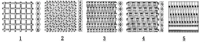
1. Plain Weave: also called tabby weave, linen weav or taffeta weave, is the most basic type of weaves.
In plain weave, the warp and weft are aligned so they form a simple criss-cross pattern. Each weft thread
crosses the warp threads by going over one, then under the next, and so on. The next weft thread goes under
the warp threads that its neighbor went over, and vice versa.
2. Twill Weave: In a twill weave, each weft or filling yarn floats across the warp yarns in a progression of interlaces
to the right or left, forming a distinct diagonal line. This diagonal line is also known as a wale. A float is the portion of
a yarn that crosses over two or more yarns from the opposite direction.
3. Plain Dutch Weave: similar with plain weave, just the weft and warp wire have different wire diameter and different mesh size.
4. Twill Dutch Weave: similar with twill weave, just the weft and warp wire have different wire diameter and different mesh size.
5. Reversed Dutch Weave: difference from standard Dutch weave lies in the thicker warp wires and less weft wires
Stainless Steel Series,Stainless Steel Wire Mesh,Stainless Steel Crimped Wire Mesh,Stainless Steel Window Screen
Anping Shengjia Hardware Mesh Co.,LTD , https://www.oilshaleshakerscreen.com